The Greatest Guide To Babyecho
The Greatest Guide To Babyecho
Blog Article
Not known Incorrect Statements About Babyecho
Table of ContentsThe Main Principles Of Babyecho The Babyecho DiariesBabyecho for DummiesThe Ultimate Guide To BabyechoIndicators on Babyecho You Need To KnowThe Babyecho DiariesNot known Details About Babyecho
:max_bytes(150000):strip_icc()/JoseLuisPelaezInc-17f79a53211940c2bc62cf23bc4185d4.jpg)
A c-section is surgery in which your infant is born with a cut that your physician makes in your tummy and womb. Regardless of what an ultrasound reveals, talk to your company about the finest look after you and your child - fetal heart doppler. Last reviewed: October, 2019
Throughout this check, they will inspect the infant is growing in the best place, whether there is more than one infant and they will certainly also examine your child's development so much. This screening is readily available between 10 14 weeks of maternity and is utilized to examine the chances of your infant being birthed with several of these conditions.
The 9-Minute Rule for Babyecho
It involves a consolidated examination of an ultrasound check and a blood test. Throughout the scan, the sonographer will measure the fluid at the rear of the child's neck to identify 'nuchal translucency' - https://dribbble.com/babydoppler1/about. They will then compute the opportunity of your infant having Down's, Edwards' or Patau's syndrome using your age, the blood test and scan outcomes
During this scan, the sonographer look for structural and developing problems in the infant. During this check consultation, you might be used testings for HIV, syphilis and liver disease B by a professional midwife. In many cases, a third-trimester scan is recommended by your midwife complying with the results of previous examinations, previous complications or existing medical problems.
The typical 2D ultrasound generates level and outlined pictures which can be made use of to see your infant's interior organs and help detect any type of internal concerns. These black and white pictures help the sonographer determine the baby's gestation, development, heartbeat, development and size. Some expectant mommies choose to have a 3D ultrasound check since they show more of a real-life photo of the infant.
9 Easy Facts About Babyecho Explained
3D ultrasound scans reveal still images of your baby's external body rather than their withins, so you can see the shape of the infant's facial functions. 4D ultrasound scans resemble 3D scans but they reveal a relocating video clip rather than still pictures. This records highlights and darkness much better, as a result producing a more clear picture of the child's face and motions.

or (8-11 weeks) (11-14 weeks) (14-18 weeks) (19-23 weeks) or (24-42 weeks) Suggested at Optional -, a lot more frequently in some conditions This check is done to and to figure out an (EDD). A is detected throughout this scan. Many parents select this check for. Is vital prior to the blood test called as (NIPT) to determine the.
Indicators on Babyecho You Should Know
Sometimes a may be called for to get and a more clear photo. This is generally performed and sometimes a may be needed. Validate that the baby's heart is present; To more accurately. This might not be needed in, where the from the is extra accurate; To; To detect whether and to analyze whether there is sharing of placenta, which will need close tracking in pregnancy; To examine the consisting of dimension of; To see if there is a low or high chance for the baby to be affected with such as Down's Disorder, Edward's Disorder and; If any, further relating to will certainly be offered at the exact same analysis by myself.
Please see below. These scans might be done, nevertheless some of the and thus, a is required to This scan is done generally at.
9 Easy Facts About Babyecho Described

Furthermore, the can be by by an. and is kept an eye on by these scans. of, andare done to get to an. around the baby is measured. and child's are checked. () The way nearer the is useful to. Sometimes, an which was in the past might be.
The Buzz on Babyecho
If, these scans might be to. on the searchings for, a may be used. During all the, a 3D scan (of the infant) can also be executed. The is dependent on the setting of the,,, amount of and. This includes, together with; This includes, along with (14-20 weeks).
A scan is vital before this test is done. If you're trying to find, prepare an examination with Dr Sankaran through her. Obstetrics & gynaecology in London.
The Of Babyecho
The test can offer valuable details, helping females and their health-care companies take care of and care for the maternity and the unborn child.
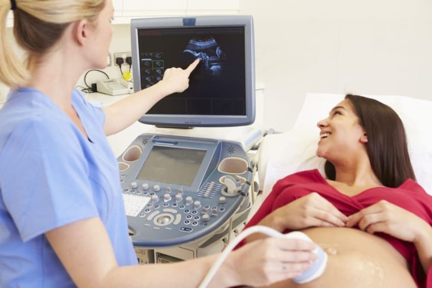
A transducer is inserted into the vagina and relaxes versus the back of the vaginal area to website here create a picture. A transvaginal ultrasound produces a sharper image and is usually utilized in very early maternity. Ultrasound machines are regarding the size of a grocery store cart. A TV display for checking out the photos is affixed to the equipment (http://prsync.com/babyecho/).
Report this page